Image Production · ARRT 2025
Pathology, Artifacts, and Processor Errors
Additive and destructive pathology, common artifacts (motion, double exposure, grid cutoff), and processor errors for the ARRT Radiography Boards.
Overview
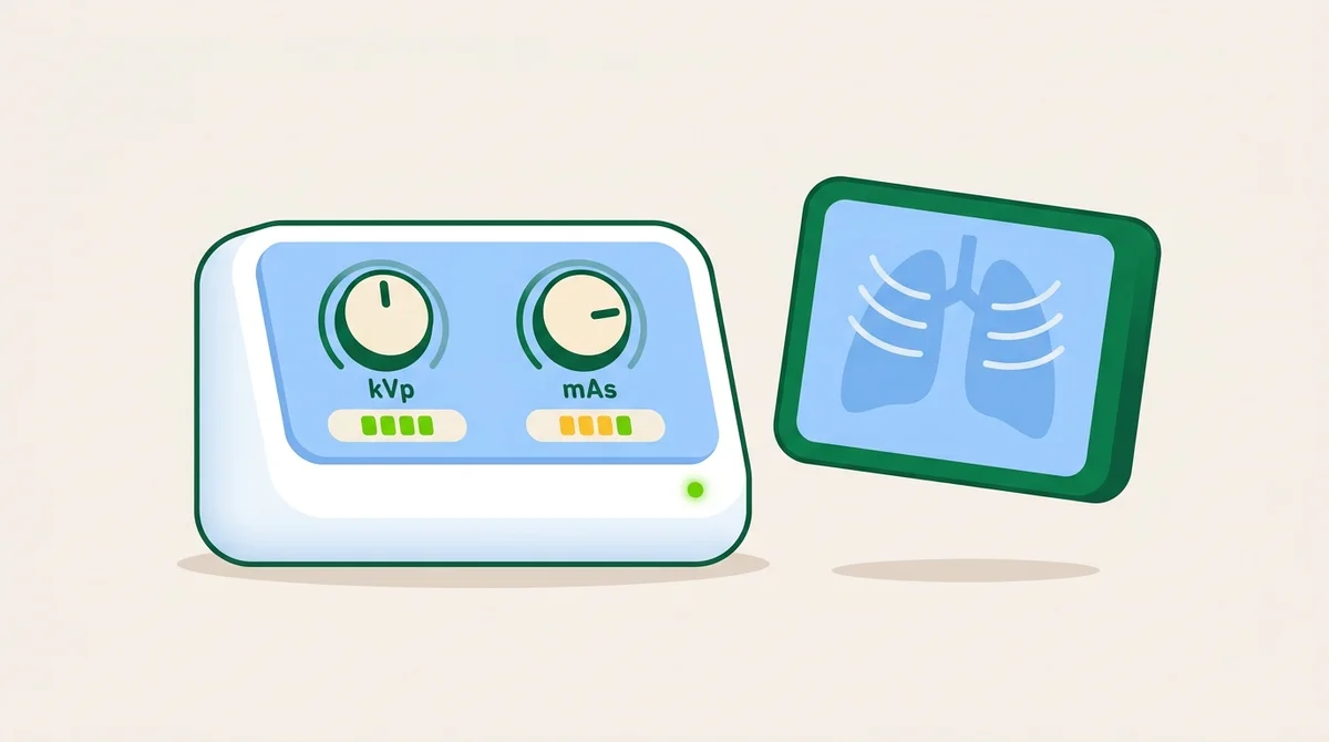
Pathology, Artifacts, and Processor Errors is a synthesis chapter, three skill sets that turn an OK radiographer into a great one. Reading the patient (does this disease ADD density or take it AWAY?), identifying the artifact (was this caused by motion, processor, or grid?), and decoding the processor error (is the image too light, too dark, or fogged?) all combine in the analytical work that follows the exposure.
Additive pathology increases tissue density and requires technique increase. Pneumonia, pleural effusion, pulmonary edema, ascites, sclerotic metastasis, and Paget's disease all demand more kVp or mAs to penetrate. Destructive pathology decreases tissue density and requires technique decrease. Pneumothorax, emphysema, osteoporosis, lytic metastasis, atrophy, and active tuberculosis all demand less mAs to avoid overexposure. The boards routinely test pairings, match the disease to the technique adjustment.
Artifacts split by source. Motion artifact: blurred edges, ghosted contours. Caused by patient or generator motion. Solution: shorter exposure time, immobilization. Double exposure: two images on one IR. Pre-CR/DR mistake. Grid cutoff: density loss across one or both edges of the image. Caused by off-center grid, off-level grid, off-focus grid, or upside-down grid. Quantum mottle: grainy, noisy image. Caused by too few photons (low mAs at high kVp). Processor errors: chemical fog (overdeveloped), low contrast (underdeveloped), pi lines (roller pressure), wet pressure (rollers carrying chemicals onto the next film). Modern CR/DR adds histogram errors, exposure-index errors, and image-processing artifacts (dead pixels, image stitching errors).
What you’ll learn in this chapter
The 13 lessons in this chapter break down as follows. The full lesson content is unlocked when you start a free account.
Reading the Patient
- The Three Categories of Image Anomalies
- Additive vs Destructive Pathology
- Spectrum of Pathological Adjustments
Identifying Artifacts
- Artifact Categories
- Exposure Artifacts: Patient Foreign Objects
- Handling Artifacts: Light, Pressure, Static
Decoding the Processor
- Chemical Imbalances
- Mechanical & Digital Artifacts
- From Identification to Prevention
Knowledge Check
- Question 1 of 4 Quiz
- Question 2 of 4 Quiz
- Question 3 of 4 Quiz
- Question 4 of 4 Quiz
Key terms in this chapter
These are the 7 terms most likely to appear on the ARRT registry from this chapter. Use them as a flashcard pre-quiz.
- Additive Pathology
- Disease that increases tissue density. Pneumonia, pleural effusion, ascites, Paget's disease. Increase technique.
- Destructive Pathology
- Disease that decreases tissue density. Pneumothorax, emphysema, osteoporosis. Decrease technique.
- Grid Cutoff
- Density loss caused by improper grid positioning (off-center, off-level, off-focus, upside down).
- Quantum Mottle
- Grainy, noisy image from too few photons reaching the IR. Caused by low mAs at high kVp.
- Motion Artifact
- Blurred edges from patient or equipment motion during exposure. Reduced by short exposure time and immobilization.
- Pi Lines
- Repeating density variations from worn or sticky processor rollers. A film-screen processor artifact.
- Exposure Index
- CR/DR metric of receptor exposure. High EI = overexposure; low EI = underexposure.
Sample practice question: Image Acquisition
One free sample from the 98-question Image Acquisition bank. See the format, the rationale style, and the difficulty before you sign up.
An adult chest x-ray is performed at 80 kVp and 4 mAs at 72 inches SID. To maintain density at 40 inches SID, what new mAs is required?
Show answer and rationale
A, Incorrect: 1 mAs would result in significant underexposure. Apply the density maintenance formula.
B, Incorrect: Distance is decreasing, so mAs should decrease, but not by half.
C, Correct: Correct. Density maintenance formula: mAs₂ = mAs₁ × (SID₂² / SID₁²) = 4 × (40² / 72²) = 4 × (1600/5184) = 4 × 0.309 ≈ 1.2 mAs. Closer distance means more intensity, so less mAs is needed.
D, Incorrect: 13 mAs would more than triple the exposure. Reversed formula error, increasing distance requires more mAs, not less.
Read the full chapter, free.
The free tier unlocks one complete chapter (13 lessons), 50 practice questions, and 1 sample timed exam. No credit card required.
Frequently asked questions
What does the ARRT Radiography Image Production category cover?
Pathology, Artifacts, and Processor Errors is a synthesis chapter, three skill sets that turn an OK radiographer into a great one. Reading the patient (does this disease ADD density or take it AWAY?), identifying the artifact (was this caused by motion, processor, or grid?), and decoding the processor error (is the image too light, too dark, or fogged?) all combine in the analytical work that follows the exposure.
How many lessons are in the Pathology, Artifacts, and Processor Errors chapter?
This chapter contains 13 lessons across 4 sections, plus a knowledge-check quiz at the end. The full lesson content is unlocked with a Premium subscription. The free tier includes the first chapter complete.
Is this chapter aligned with the ARRT 2025 Content Specifications?
Yes. Every chapter on this site maps directly to the ARRT Radiography Content Specifications effective 2025. This chapter falls under the Image Production domain of the official ARRT exam blueprint.