Radiographic Procedures · ARRT 2025
Myelography, Cardiac Cath, Angiography, Venography for the ARRT
Myelography, cardiac catheterization, aortography, angiography, and venography techniques for the ARRT Radiography Boards.
Overview
Special Procedures covers the imaging studies of the central nervous system and the circulatory system. These exams are typically performed in fluoroscopic suites or angiographic labs and involve catheter or needle placement, contrast injection, and dynamic imaging. The ARRT registry expects familiarity with the names, the access sites, and the safety concerns, not the procedural skill itself.
Myelography is contrast-enhanced imaging of the spinal subarachnoid space. Lumbar puncture below L3 (to avoid the spinal cord, which ends at L1–L2 in adults), water-soluble iodinated contrast injection, then fluoroscopic positioning to demonstrate cervical, thoracic, or lumbar pathology. The patient is tilted on the fluoroscopic table to flow contrast cephalad or caudad as needed. Post-procedure: head-of-bed elevated to keep contrast away from the brain.
Cardiac catheterization: percutaneous femoral or radial artery access, catheter advanced retrograde to the coronary ostia, contrast injection during cine imaging. Coronary angiography demonstrates LAD, circumflex, and right coronary artery anatomy. Aortography: thoracic or abdominal aorta evaluation via translumbar or femoral approach. Cerebral angiography: imaging of the carotid and vertebral circulation, performed for aneurysm, AVM, or stroke workup. Peripheral angiography evaluates upper or lower extremity arterial disease. Venography images the venous system, the most common indication is suspected DVT, though Doppler ultrasound has largely supplanted contrast venography for that purpose. Across all special procedures, radiation safety is paramount: the patient is on the table for extended fluoroscopic time, and the radiographer/operator must use lead, distance, and dose-area-product monitoring.
What you’ll learn in this chapter
The 11 lessons in this chapter break down as follows. The full lesson content is unlocked when you start a free account.
Central Nervous System
- CNS Anatomy: Atlas of Command
- Myelography: Visualizing the Spinal Canal
- Myelography Contrast: Water-Soluble vs Legacy
Circulatory System
- The Four-Chambered Heart
- The Aorta & Its Branches
- Lower Extremity Venography
- Significant Conditions: CNS & Circulatory
Knowledge Check
- Question 1 of 4 Quiz
- Question 2 of 4 Quiz
- Question 3 of 4 Quiz
- Question 4 of 4 Quiz
Key terms in this chapter
These are the 7 terms most likely to appear on the ARRT registry from this chapter. Use them as a flashcard pre-quiz.
- Myelography
- Contrast study of the spinal subarachnoid space. Lumbar puncture below L3, water-soluble iodinated contrast.
- Cardiac Catheterization
- Catheter-based imaging of coronary arteries via femoral or radial artery access. Cine imaging during contrast injection.
- Coronary Angiography
- Imaging of the LAD, circumflex, and right coronary arteries. Performed during cardiac cath.
- Cerebral Angiography
- Contrast study of carotid and vertebral arteries. Used for aneurysm, AVM, and acute stroke workup.
- Aortography
- Contrast imaging of the thoracic or abdominal aorta. Femoral or translumbar approach.
- Venography
- Contrast imaging of veins. Largely replaced by Doppler ultrasound for DVT diagnosis.
- DSA (Digital Subtraction Angiography)
- Subtraction technique that removes overlying bone and tissue, leaving only the contrast-filled vessels.
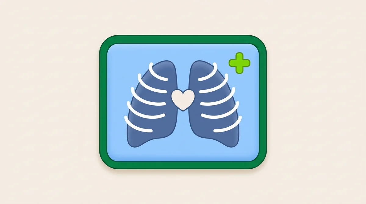
Sample practice question: Thorax and Abdomen
One free sample from the 75-question Thorax and Abdomen bank. See the format, the rationale style, and the difficulty before you sign up.
On a properly positioned and exposed PA chest radiograph in an adult, how many posterior ribs should be visible above the diaphragm to indicate adequate inspiration?
Show answer and rationale
A, Incorrect: 8 posterior ribs typically indicates underinspiration. Repeat with full inspiration.
B, Correct: Correct. The standard is at least 10 posterior ribs visible above the diaphragm on a properly inspired adult PA chest. Underinspiration (only 8 visible) compresses lung markings and may mimic pulmonary disease.
C, Incorrect: Counting anterior ribs is less reliable because they're more variable and angle differently. Use posterior ribs as the standard.
D, Incorrect: Costophrenic angle visibility is part of quality assessment but does not specifically indicate inspiration adequacy.
Read the full chapter, free.
The free tier unlocks one complete chapter (11 lessons), 50 practice questions, and 1 sample timed exam. No credit card required.
Frequently asked questions
What does the ARRT Radiography Radiographic Procedures category cover?
Special Procedures covers the imaging studies of the central nervous system and the circulatory system. These exams are typically performed in fluoroscopic suites or angiographic labs and involve catheter or needle placement, contrast injection, and dynamic imaging. The ARRT registry expects familiarity with the names, the access sites, and the safety concerns, not the procedural skill itself.
How many lessons are in the Myelography, Cardiac Cath, Angiography, Venography for the ARRT chapter?
This chapter contains 11 lessons across 3 sections, plus a knowledge-check quiz at the end. The full lesson content is unlocked with a Premium subscription. The free tier includes the first chapter complete.
Is this chapter aligned with the ARRT 2025 Content Specifications?
Yes. Every chapter on this site maps directly to the ARRT Radiography Content Specifications effective 2025. This chapter falls under the Radiographic Procedures domain of the official ARRT exam blueprint.