Radiographic Procedures · ARRT 2025
Hand, Wrist & Forearm Positioning for the ARRT Exam
27 bones, the carpal mnemonic, PIP and MCP centering points, scaphoid and ulnar deviation views, Gaynor-Hart carpal canal, and named fractures for the ARRT exam.
Overview
Hand, Wrist and Forearm covers 27 bones per side: 14 phalanges, 5 metacarpals and 8 carpals in two rows of four, memorized with the mnemonic Send Letter To Peter To Tell 'em Come Home. The scaphoid is the most frequently fractured carpal, which is why the wrist has an entire family of special scaphoid projections and why the ARRT keeps asking about them.
Most positioning questions here are really centering questions. Fingers center at the PIP joint, the thumb at the first MCP joint, the PA and oblique hand at the third MCP, the fan lateral at the second MCP, the wrist at the midcarpal area, and the forearm at its midpoint with both joints on the image. The forearm is never radiographed pronated, because pronation crosses the radius over the ulna near the upper third.
Four central ray angles carry the special views: 10 to 15 degrees proximally for the ulnar deviation scaphoid projection, 15 degrees for the modified Robert's method aimed at Bennett's fracture, 25 to 30 degrees for the Gaynor-Hart carpal canal, and 45 degrees for the carpal bridge. The deviation logic is one sentence: ulnar deviation opens the radial-side carpals, radial deviation opens the ulnar side.
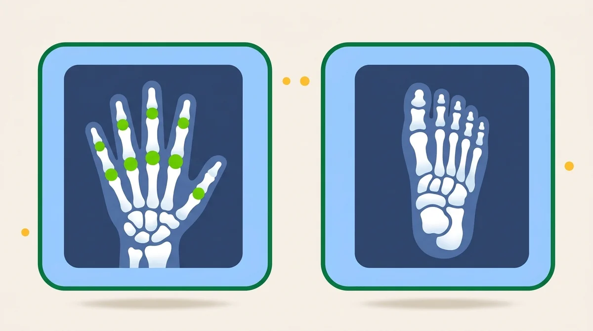
The named injuries are a location plus a displacement. A Colles fracture is a distal radius fracture displaced posteriorly, Smith's is its anterior reverse, a boxer's fracture crosses the fifth metacarpal neck, and skier's thumb is a torn ulnar collateral ligament imaged with the Folio stress method. The Norgaard ball-catcher view hunts early rheumatoid arthritis at the MCP and PIP joints, with both hands compared on a single exposure.
What you’ll learn in this chapter
The 33 lessons in this chapter break down as follows. The full lesson content is unlocked when you start a free account.
Procedures
- Hand, Wrist & Forearm
Hand anatomy
- Phalanges and Metacarpals
Joints
- Every Joint, Named and Classified
- Quiz
Carpals
- The Eight Carpal Bones
- Quiz
Wrist anatomy
- The Wrist Joint, Canal and Fat Stripes
Forearm anatomy
- Radius and Ulna
Technique
- Technical Factors for the Upper Limb
Digits
- Fingers, Digits 2 to 5
Thumb
- The Thumb Routine
- Modified Robert's, Lewis and Folio
- Quiz
Hand
- The Hand Routine
- Lateral Alternates and the Ball-Catcher
- Quiz
Wrist
- The Wrist Routine
Scaphoid
- Scaphoid, Ulnar and Radial Deviation
- Quiz
Carpal canal
- Gaynor-Hart and the Carpal Bridge
- Quiz
Forearm
- The Forearm
- Quiz
Image critique
- Critiquing a Hand or Wrist Image
Clinical indications
- Named Fractures and Pathology
Exam traps
- Answering the Outline Directly
- Quiz
Recap
- What to Carry Out of This Chapter
- Quiz
- Quiz
- Quiz
- Quiz
- Quiz
Key terms in this chapter
These are the 8 terms most likely to appear on the ARRT registry from this chapter. Use them as a flashcard pre-quiz.
- Scaphoid
- The largest bone of the proximal carpal row and the most frequently fractured carpal. Located 2 cm distal and medial to the radial styloid process.
- Carpal Sulcus
- The concave palmar surface of the carpals, also called the carpal tunnel, carrying the median nerve and tendons. Imaged tangentially with the Gaynor-Hart method.
- Ulnar Deviation
- Bending the wrist toward the ulnar side, which opens the radial-side carpals: scaphoid, trapezium and trapezoid. Commonly called the scaphoid projection.
- Modified Robert's Method
- An AP axial thumb projection with the CR angled 15 degrees proximally to the first CMC joint, used to rule out a Bennett's fracture.
- Gaynor-Hart Method
- The tangential carpal canal projection: wrist hyperextended, CR angled 25 to 30 degrees to the long axis of the hand, showing the pisiform and hamulus in profile.
- Norgaard Method
- The ball-catcher's position, both hands supinated and rotated 45 degrees on one exposure, used to find early rheumatoid arthritis at the MCP and PIP joints.
- Colles Fracture
- A transverse fracture of the distal radius with posterior displacement of the distal fragment. An ulnar styloid fracture accompanies it in 50 to 60 percent of cases.
- Bennett's Fracture
- A fracture of the base of the first metacarpal extending into the CMC joint, with subluxation and some posterior displacement.
Sample practice question: Extremity
One free sample from the 105-question Extremity bank. See the format, the rationale style, and the difficulty before you sign up.
A patient presents with wrist pain after falling on an outstretched hand. The PA, lateral, and oblique wrist projections appear normal but clinical suspicion for scaphoid fracture remains. What additional projection should be performed?
Show answer and rationale
A, Incorrect: The carpal tunnel projection demonstrates the carpal tunnel itself, not the scaphoid in particular.
B, Correct: Correct. The Stecher method (PA wrist with ulnar deviation, central ray angled 20° proximally toward the elbow) elongates the scaphoid and reveals fractures missed on routine PA. It is the standard supplementary view for suspected scaphoid fracture.
C, Incorrect: AP oblique medial rotation is for the elbow, not the wrist. Wrist obliques are routinely lateral rotation.
D, Incorrect: Radial deviation does not elongate the scaphoid. Ulnar deviation is required for the Stecher.
Hands-on
Practice the positioning
Open the Positioning Lab to drill body position, central ray, anatomy, and common errors for each projection in this chapter.
- Elbow AP Seated, arm extended on IR, palm supinated. SID 40".
- Elbow Lateral Seated, elbow flexed 80-90 degrees, hand in lateral position. SID 40".
- Elbow AP Oblique (Lateral Rotation) Arm extended, rotated 45 degrees laterally until 1st/2nd digits touch the table.
- Elbow AP Oblique (Medial Rotation) Arm extended, hand internally rotated 45 degrees from true AP.
- Elbow Axiolateral, Coyle (Radial Head) Elbow flexed 90 degrees, palm flat on IR. CR angled 45 degrees toward shoulder.
- Elbow Axiolateral, Coyle (Coronoid Process) Elbow flexed 80 degrees, palm flat on IR. CR angled 45 degrees away from shoulder.
- Thumb AP Seated, thumb abducted at 45 degrees on IR. SID 40".
- Thumb PA Oblique Seated, hand pronated, thumb obliqued 45 degrees. SID 40".
- Thumb Lateral Seated, thumb abducted, medial surface perpendicular. SID 40".
- Digits 2-5 PA Seated, hand pronated, palm down on IR. SID 40".
- Digits 2-5 PA Oblique Seated, hand rotated 45 degrees, ulnar side raised. SID 40".
- Digits 2-5 Lateral Seated, medial side on IR, fingers extended. SID 40".
- Hand PA Seated with forearm on table, palm down. SID 40".
- Hand PA Oblique Seated with hand obliqued 45 degrees, ulnar side raised. SID 40".
- Hand Lateral Seated with medial side of hand on IR. SID 40".
- Wrist PA Seated with forearm on table, wrist pronated. SID 40".
- Wrist PA Oblique Seated with wrist rotated 45 degrees, ulnar side raised. SID 40".
- Wrist Lateral Seated with elbow flexed 90 degrees, wrist lateral. SID 40".
- Wrist PA in Ulnar Deviation Seated, forearm extended on IR, hand deviated toward ulnar side. SID 40".
- Wrist PA Axial for Scaphoid Seated, hand in mild ulnar deviation. CR angled 15–20° toward elbow.
- Wrist Tangential (Carpal Canal) Seated, wrist hyperextended. CR angled 25–30° to long axis of hand.
- Forearm AP Seated, forearm extended on IR, palm supinated. SID 40".
- Forearm Lateral Seated, forearm lateral on IR with thumb up. SID 40".
- Humerus AP Upright, arm at side with elbow flexed 90 degrees, palm inward. SID 40".
- Humerus Lateral (Upright) Upright, arm internally rotated, palm backward. SID 40".
- Humerus Lateral (Recumbent) Supine, arm internally rotated, palm down. SID 40".
- Shoulder AP Erect, arm at side with slight external rotation. SID 40".
- Shoulder AP Oblique, Grashey Method Body rotated 35-45 degrees. CR perpendicular to scapular plane. SID 40".
- Shoulder Scapular Y Lateral or oblique erect position, arm abducted. CR perpendicular to scapular Y. SID 40".
- Shoulder Transthoracic Lateral Lateral erect, affected shoulder against IR. Breathing technique. SID 40".
- AC Joints AP Erect, bilateral AC joints. Arms at sides, weights attached for separation. SID 72".
- AC Joints AP with Weights Erect, bilateral AC joints. 5 to 10 lb weights suspended from both wrists. SID 72".
- Clavicle AP Erect, arm at side. CR perpendicular to clavicle midshaft. SID 40". Include entire clavicle.
- Clavicle AP Axial Erect, CR 15-30 degrees cephalad. Clavicle clear of ribs. SID 40".
- Scapula AP Erect, arm abducted 90 degrees. CR perpendicular to midscapula. SID 40".
- Scapula Lateral Lateral erect, 45-60 degree rotation. CR perpendicular to scapular body. SID 40".
Read 3 chapters free.
The free tier unlocks 3 complete chapters, 50 practice questions, and 1 sample timed exam. No credit card required.
Frequently asked questions
What does the ARRT Radiography Radiographic Procedures category cover?
Hand, Wrist and Forearm covers 27 bones per side: 14 phalanges, 5 metacarpals and 8 carpals in two rows of four, memorized with the mnemonic Send Letter To Peter To Tell 'em Come Home. The scaphoid is the most frequently fractured carpal, which is why the wrist has an entire family of special scaphoid projections and why the ARRT keeps asking about them.
How many lessons are in the Hand, Wrist & Forearm Positioning for the ARRT Exam chapter?
This chapter contains 33 lessons across 18 sections, plus a knowledge-check quiz at the end. The full lesson content is unlocked with a Premium subscription. The free tier includes 3 complete chapters.
Is this chapter aligned with the ARRT 2025 Content Specifications?
Yes. Every chapter on this site maps directly to the ARRT Radiography Content Specifications effective 2025. This chapter falls under the Radiographic Procedures domain of the official ARRT exam blueprint.