Procedures · ARRT Radiography
Foot, Ankle & Calcaneus Positioning for the ARRT Exam
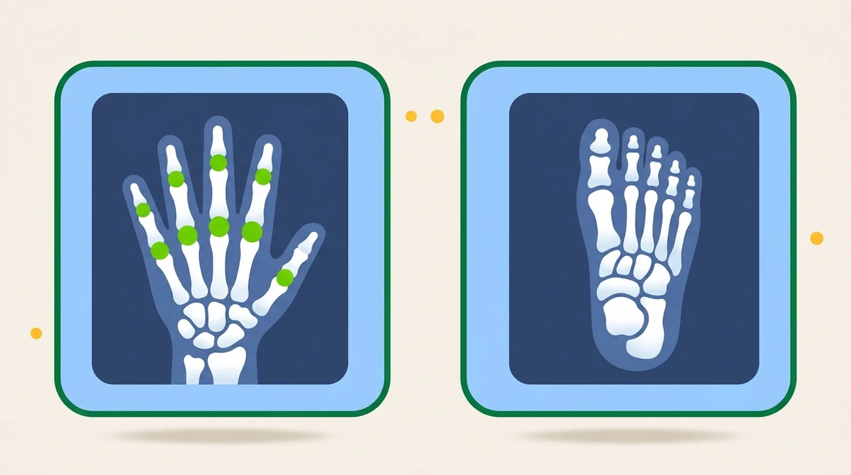
The 26 foot bones, the three frontal ankle rotations (0, 15-20, and 45 degrees), the 40 degree axial calcaneus, and weight-bearing foot projections for the ARRT Radiography exam.
Overview
Foot, Ankle and Calcaneus covers everything at or below the ankle joint: toes, foot, sesamoids, calcaneus and the ankle itself. It sits inside the ARRT Procedures domain, worth 66 of the 200 scored questions until 28 February 2027 and 72 from 1 March 2027. One foot contains 26 bones: 14 phalanges, 5 metatarsals and 7 tarsals.
The AP ankle is taken at 0 degrees, the AP mortise at 15 to 20 degrees of internal rotation, and the AP oblique at 45 degrees. The small rotation opens the entire mortise, with an even 3 to 4 mm space over the talar surface, while the 45 degree oblique opens the distal tibiofibular joint, while the medial malleolus and talus partly superimpose. Four more angles carry marks: 10 degrees posteriorly on the AP foot, 10 to 15 degrees on the AP toes, 15 degrees on the weight-bearing AP foot and 40 degrees cephalad on the plantodorsal axial calcaneus.
Centering repeats: the base of the third metatarsal serves the AP and oblique foot, a point midway between the malleoli serves four of the five ankle projections, and the lateral calcaneus centers 1 inch below the medial malleolus. Rotation checks are single landmarks: the distal fibula over the posterior half of the tibia on any lateral, and the sustentaculum tali in profile on the axial calcaneus.
A Lisfranc injury needs weight-bearing AP and lateral views plus a 30 degree medial oblique, gout classically strikes the first MTP joint, and Bontrager's cast conversions add 5 to 7 kV for a small to medium plaster cast, 8 to 10 kV for a large plaster cast, or 3 to 4 kV for fiberglass. On AP stress views, a physician or other health professional holds or straps the foot and ankle, or the patient holds the position with long gauze looped around the ball of the foot (Bontrager).
What you’ll learn in this chapter
The 20 study slides and 15 quiz questions in this chapter break down as follows. 3 chapters are free in full with a free account. The others come with a paid plan, and paid plans open soon.
Foot & Ankle
- Foot, Ankle & Calcaneus
Anatomy
- Phalanges and Metatarsals
- The Seven Tarsals
- Quiz
- Arches, Surfaces and Motions
- Quiz
Ankle anatomy
- The Ankle Joint
- Quiz
Technique
- The Lower Limb Technique Baseline
Toes
- The Toes
- Tangential Sesamoids
- Quiz
Foot
- The Foot, Six Projections
- AP Foot
- Quiz
- Oblique Foot
- Quiz
- Lateral and Weight-Bearing Foot
Calcaneus
- The Calcaneus
- Quiz
Ankle
- The Ankle, Five Standard Projections
- AP Ankle and AP Stress
- Mortise Against 45° Oblique
- Quiz
- Lateral Ankle
- Quiz
Critique
- Image Critique
- Quiz
Clinical indications
- Clinical Indications
Recap
- What to Carry Out of This Chapter
- Quiz
- Quiz
- Quiz
- Quiz
- Quiz
Key terms in this chapter
These are 7 key terms from this chapter. Use them as a flashcard pre-quiz.
- Ankle Mortise
- The three-sided socket formed by the tibial plafond, medial malleolus and lateral malleolus, into which the talus fits. A 15 to 20 degree internal rotation opens it, showing an even 3 to 4 mm space over the talar surface.
- Tarsals
- The seven bones of the proximal foot: calcaneus, talus, cuboid, navicular and three cuneiforms. The talus is the only tarsal that is actually part of the ankle joint.
- Sustentaculum Tali
- A shelf on the medial proximal calcaneus that supports the talus. A portion of it seen in profile medially is the entire rotation check on the axial calcaneus.
- Plantodorsal Axial Calcaneus
- Routine heel projection with the CR angled 40 degrees cephalad, entering at the base of the third metatarsal. With analog imaging, the dense calcaneus takes 8 to 10 kV more than the other foot projections; the digital range is 70 ± 5 kV.
- AP Foot (Dorsoplantar)
- Routine foot projection with the CR angled 10 degrees posteriorly toward the heel, centered to the base of the third metatarsal. A foreign body study drops the angle and uses a perpendicular ray.
- Lisfranc Joint Injury
- Separation or avulsion between the bases of the first and second metatarsals and the cuneiforms. Worked up with weight-bearing AP and lateral projections plus a 30 degree medial oblique, because recumbent films can look normal.
- AP Stress Views
- Inversion and eversion ankle projections that demonstrate joint separation after a ligament tear. A physician or other health professional holds or straps the foot and ankle, or the patient holds it with long gauze.
Sample practice question: Extremity
One free sample question from the Extremity subcategory, like those in its 377-question bank. Several chapters share each sample, so it may not match this chapter's topic. See the format, the rationale style, and the difficulty before you sign up.
A patient presents with wrist pain after falling on an outstretched hand. The PA, lateral, and oblique wrist projections appear normal but clinical suspicion for scaphoid fracture remains. What additional projection should be performed?
Show answer and rationale
A, Incorrect: The carpal tunnel projection demonstrates the carpal tunnel itself.
B, Correct: Correct. ARRT and Merrill's call the Stecher method a PA axial scaphoid projection, while Bontrager titles its modified version a PA scaphoid. Both books raise the hand end 20° with a perpendicular CR, and Bontrager adds ulnar deviation. Merrill's also gives a variation with the CR 20° toward the elbow, and Bontrager gives a separate ulnar-deviation method with the CR angled 10° to 15° proximally. Use your program's text. Bontrager notes nondisplaced fractures may still need more projections or CT.
C, Incorrect: This is a real wrist projection, but it shows the medial carpals, not the scaphoid.
D, Incorrect: This is not a scaphoid projection. Radial deviation is its own PA wrist projection, and it shows the medial carpals.
Hands-on
Practice the positioning
Open the Positioning Lab to drill body position, central ray, anatomy, and common errors for each projection in this chapter.
- Toes AP Foot flat on IR, CR 10-15 degrees toward the heel to the MTP joint in question. SID 40".
- Toes Lateral Affected toe in true lateral on IR, CR perpendicular. SID 40".
- Toes AP Oblique Foot rotated 30-45 degrees, medially for toes 1-3 and laterally for toes 4-5. CR perpendicular. SID 40".
- Sesamoids Tangential, Lewis Method Patient prone, toe flexed back on IR. CR perpendicular and tangential. SID 40".
- Foot AP Axial Sole flat on IR, CR angled 10° cephalad toward heel. SID 40".
- Foot Medial Oblique Foot rotated medially 30-40 degrees (Bontrager) or 30 (Merrill's), CR perpendicular. SID 40".
- Calcaneus Axial (Plantodorsal) Foot dorsiflexed, CR angled 40° cephalad through plantar surface. SID 40".
- Calcaneus Lateral (Mediolateral) Patient lateral recumbent on affected side, CR perpendicular to calcaneus. SID 40".
- Ankle AP Supine, leg extended and not rotated, CR perpendicular to the ankle joint. SID 40".
- Ankle Lateral (Mediolateral) Patient lateral recumbent on affected side, CR perpendicular to ankle. SID 40".
- Ankle Lateral (Lateromedial) Patient lateral on non-affected side, medial ankle on IR. CR perpendicular. SID 40".
- AP Medial Oblique Ankle Supine, ankle internally rotated 45 degrees. SID 40".
- AP Medial Oblique Mortise Ankle Supine, leg internally rotated 15-20°. Mortise open. SID 40".
- AP Weight-Bearing Ankle Upright on a low platform, heels against the IR, toes forward. SID 40".
Read 3 chapters free.
The free tier unlocks 3 complete chapters, 50 practice questions, and 1 sample timed exam. No credit card required.
Frequently asked questions
What does the Foot, Ankle & Calcaneus Positioning for the ARRT Exam chapter cover?
Foot, Ankle and Calcaneus covers everything at or below the ankle joint: toes, foot, sesamoids, calcaneus and the ankle itself. It sits inside the ARRT Procedures domain, worth 66 of the 200 scored questions until 28 February 2027 and 72 from 1 March 2027. One foot contains 26 bones: 14 phalanges, 5 metatarsals and 7 tarsals.
How many study slides are in the Foot, Ankle & Calcaneus Positioning for the ARRT Exam chapter?
This chapter has 20 study slides and 15 quiz questions across 11 sections. The full chapter comes with a paid plan, and paid plans open soon. The free tier includes 3 complete chapters.
Is this chapter aligned with the current ARRT Content Specifications?
Every chapter on this site is mapped to the ARRT Radiography Content Specifications, but our line-by-line check against ARRT's outline isn't finished, so keep ARRT's outline beside it. Nothing here follows your exam date, so check which version applies to you. This chapter falls under the Procedures domain of the official ARRT exam blueprint.