Radiographic Procedures · ARRT 2025
Trauma Radiography for ARRT
Trauma protocols, fracture terminology, the Don't Move Rule, and adaptive projections (cross-table lateral, Danelius-Miller) for the ARRT Radiography Boards.
Overview
Trauma and Adaptive Positioning answers a single clinical question: when the patient cannot move into the routine position, what do you do instead? The ARRT registry tests this material in scenario format, given an unstable fracture, a comatose patient, or a patient on a backboard, identify the correct projection without making the injury worse.
The Don't Move Rule governs trauma imaging: never move the patient or the affected part. Move the tube and the IR instead. For a suspected hip fracture, do not abduct or internally rotate the leg, use the Danelius-Miller cross-table axiolateral. For a suspected C-spine injury, do not lift the head, use a horizontal-beam cross-table lateral first to clear the cervical spine, then proceed. For unstable forearm fractures where the patient cannot pronate or supinate, take two AP projections (one for the humerus side, one for the radius/ulna side) rather than forcing rotation.
Fracture terminology is heavily tested. Open (compound) versus closed (simple). Complete versus incomplete (greenstick). Displaced versus non-displaced. Transverse, oblique, spiral, comminuted, segmental. Specific named patterns: Salter-Harris (pediatric epiphyseal), pathologic (through diseased bone), stress, avulsion, impacted. Healing-stage terms: callus, bridging, malunion, nonunion. The chapter also covers the radiographer's documented refusal, when an ordered projection violates the Don't Move Rule, you must document why a substitute view was performed and which radiologist authorized it.
What you’ll learn in this chapter
The 7 lessons in this chapter break down as follows. The full lesson content is unlocked when you start a free account.
The Language of Fractures
- Fracture Terminology
Clinical Judgment: When NOT to Position
- Suspected Hip Fracture: Never Rotate
- Suspected Patellar Fracture: Don’t Flex
Knowledge Check
- Question 1 of 4 Quiz
- Question 2 of 4 Quiz
- Question 3 of 4 Quiz
- Question 4 of 4 Quiz
Key terms in this chapter
These are the 7 terms most likely to appear on the ARRT registry from this chapter. Use them as a flashcard pre-quiz.
- Don't Move Rule
- In trauma, move the tube and IR, never the patient or the injured part. Adapt the projection, not the anatomy.
- Cross-Table Lateral
- Lateral projection performed with a horizontal central ray and the patient supine. Used for trauma C-spine and hip imaging.
- Salter-Harris Fracture
- Pediatric fracture involving the epiphyseal plate. Classified Types I–V by the Salter-Harris system.
- Compound Fracture
- Fracture in which the bone breaks through the skin (open). Distinct from a closed/simple fracture.
- Comminuted Fracture
- Fracture in which the bone is broken into three or more fragments.
- Greenstick Fracture
- Incomplete fracture seen in pediatric bones. The cortex bends and breaks on one side only.
- Avulsion Fracture
- A small piece of bone is pulled away by the attached ligament or tendon. Common at insertions.
Sample practice question: Extremity
One free sample from the 105-question Extremity bank. See the format, the rationale style, and the difficulty before you sign up.
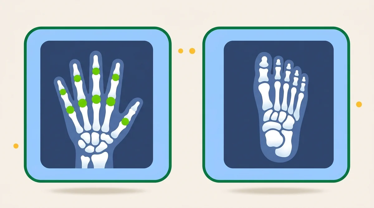
A patient presents with wrist pain after falling on an outstretched hand. The PA, lateral, and oblique wrist projections appear normal but clinical suspicion for scaphoid fracture remains. What additional projection should be performed?
Show answer and rationale
A, Incorrect: The carpal tunnel projection demonstrates the carpal tunnel itself, not the scaphoid in particular.
B, Correct: Correct. The Stecher method (PA wrist with ulnar deviation, central ray angled 20° proximally toward the elbow) elongates the scaphoid and reveals fractures missed on routine PA. It is the standard supplementary view for suspected scaphoid fracture.
C, Incorrect: AP oblique medial rotation is for the elbow, not the wrist. Wrist obliques are routinely lateral rotation.
D, Incorrect: Radial deviation does not elongate the scaphoid. Ulnar deviation is required for the Stecher.
Read the full chapter, free.
The free tier unlocks one complete chapter (7 lessons), 50 practice questions, and 1 sample timed exam. No credit card required.
Frequently asked questions
What does the ARRT Radiography Radiographic Procedures category cover?
Trauma and Adaptive Positioning answers a single clinical question: when the patient cannot move into the routine position, what do you do instead? The ARRT registry tests this material in scenario format, given an unstable fracture, a comatose patient, or a patient on a backboard, identify the correct projection without making the injury worse.
How many lessons are in the Trauma Radiography for ARRT chapter?
This chapter contains 7 lessons across 3 sections, plus a knowledge-check quiz at the end. The full lesson content is unlocked with a Premium subscription. The free tier includes the first chapter complete.
Is this chapter aligned with the ARRT 2025 Content Specifications?
Yes. Every chapter on this site maps directly to the ARRT Radiography Content Specifications effective 2025. This chapter falls under the Radiographic Procedures domain of the official ARRT exam blueprint.