Radiographic Procedures · ARRT 2025
Upper Extremity Positioning
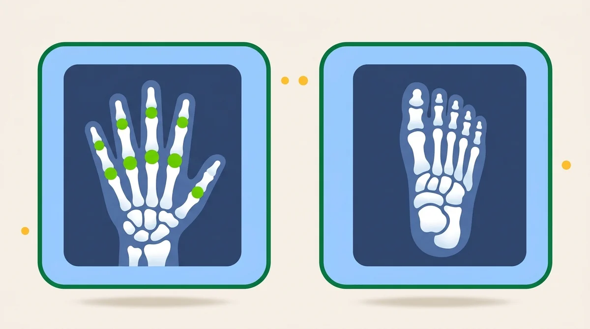
Standard projections for the hand, wrist, elbow, humerus, shoulder, and clavicle. Routine views, special projections (Stecher, scaphoid, AP axial clavicle), and pathology for the ARRT exam.
Overview
Upper Extremity is one of the highest-yield chapters on the ARRT Radiography registry. Roughly one in eight registry questions tests upper-extremity anatomy or projection. The chapter is organized distally to proximally (fingers → hand → wrist → forearm → elbow → humerus → shoulder → clavicle) and the workflow follows the same logic clinically.
The routine hand series is PA, PA oblique (45° lateral rotation), and lateral. The wrist series is PA, lateral, and PA oblique (45° lateral rotation). For suspected scaphoid (navicular) fracture, add the Stecher (PA with ulnar deviation, central ray angled 20° proximally toward the elbow), it elongates the scaphoid and reveals fractures missed on routine PA. The elbow routine is AP, internal oblique, external oblique, and lateral. When the patient cannot fully extend, two AP projections are required (one for the humerus, one for the forearm) per the trauma protocol.
Shoulder routines split by clinical question. For suspected dislocation, the standard is AP internal rotation and AP external rotation, plus a transthoracic lateral or scapular Y. For suspected AC joint separation, weighted and unweighted bilateral AP projections. The Grashey (AP oblique, 35–45° rotation toward the affected side) shows the glenohumeral joint space in profile. Common pathology covered on the boards: Colles fracture (distal radius), boxer's fracture (5th metacarpal neck), Bennett fracture (1st metacarpal base), Smith fracture, scaphoid fracture, Monteggia and Galeazzi fractures of the forearm.
What you’ll learn in this chapter
The 17 lessons in this chapter break down as follows. The full lesson content is unlocked when you start a free account.
Skeletal Foundations
- Functions of the Skeletal System
- Anatomy of a Long Bone
- Articulations: How Bones Connect
Hand & Wrist
- Anatomy of the Hand
- The Eight Carpal Bones
- Common Hand & Wrist Pathologies
Forearm & Elbow
- Forearm Anatomy: Radius & Ulna
- The Elbow Joint
- Forearm & Elbow Projections
Humerus & Shoulder
- The Humerus
- The Shoulder Girdle
- Common Shoulder Injuries
- Shoulder Projections
Knowledge Check
- Question 1 of 4 Quiz
- Question 2 of 4 Quiz
- Question 3 of 4 Quiz
- Question 4 of 4 Quiz
Key terms in this chapter
These are the 7 terms most likely to appear on the ARRT registry from this chapter. Use them as a flashcard pre-quiz.
- Stecher Method
- PA wrist projection with ulnar deviation and 20° proximal central-ray angle. Elongates the scaphoid to reveal fractures.
- Grashey Method
- AP oblique shoulder with 35–45° body rotation toward the affected side. Demonstrates the glenohumeral joint space in profile.
- Colles Fracture
- Transverse fracture of the distal radius with dorsal (posterior) displacement of the distal fragment. From a fall on outstretched hand.
- Boxer's Fracture
- Fracture of the 5th metacarpal neck. Common from punching with a closed fist.
- Scaphoid Fracture
- Most commonly fractured carpal bone. High risk of avascular necrosis. Often missed on routine PA, use the Stecher.
- AC Joint Separation
- Acromioclavicular ligament injury. Imaged with weighted and unweighted bilateral AP projections of the shoulders.
- Bennett Fracture
- Fracture-dislocation at the base of the first metacarpal. Often unstable, requires orthopedic management.
Sample practice question: Extremity
One free sample from the 105-question Extremity bank. See the format, the rationale style, and the difficulty before you sign up.
A patient presents with wrist pain after falling on an outstretched hand. The PA, lateral, and oblique wrist projections appear normal but clinical suspicion for scaphoid fracture remains. What additional projection should be performed?
Show answer and rationale
A, Incorrect: The carpal tunnel projection demonstrates the carpal tunnel itself, not the scaphoid in particular.
B, Correct: Correct. The Stecher method (PA wrist with ulnar deviation, central ray angled 20° proximally toward the elbow) elongates the scaphoid and reveals fractures missed on routine PA. It is the standard supplementary view for suspected scaphoid fracture.
C, Incorrect: AP oblique medial rotation is for the elbow, not the wrist. Wrist obliques are routinely lateral rotation.
D, Incorrect: Radial deviation does not elongate the scaphoid. Ulnar deviation is required for the Stecher.
Hands-on
Practice the positioning
Open the Positioning Lab to drill body position, central ray, anatomy, and common errors for each projection in this chapter.
- Elbow AP Seated, arm extended on IR, palm supinated. SID 40".
- Elbow Lateral Seated, elbow flexed 80-90 degrees, hand in lateral position. SID 40".
- Elbow AP Oblique (Lateral Rotation) Arm extended, rotated 45 degrees laterally until 1st/2nd digits touch the table.
- Elbow AP Oblique (Medial Rotation) Arm extended, hand internally rotated 45 degrees from true AP.
- Elbow Axiolateral, Coyle (Radial Head) Elbow flexed 90 degrees, palm flat on IR. CR angled 45 degrees toward shoulder.
- Elbow Axiolateral, Coyle (Coronoid Process) Elbow flexed 80 degrees, palm flat on IR. CR angled 45 degrees away from shoulder.
- Thumb AP Seated, thumb abducted at 45 degrees on IR. SID 40".
- Thumb PA Oblique Seated, hand pronated, thumb obliqued 45 degrees. SID 40".
- Thumb Lateral Seated, thumb abducted, medial surface perpendicular. SID 40".
- Digits 2-5 PA Seated, hand pronated, palm down on IR. SID 40".
- Digits 2-5 PA Oblique Seated, hand rotated 45 degrees, ulnar side raised. SID 40".
- Digits 2-5 Lateral Seated, medial side on IR, fingers extended. SID 40".
- Hand PA Seated with forearm on table, palm down. SID 40".
- Hand PA Oblique Seated with hand obliqued 45 degrees, ulnar side raised. SID 40".
- Hand Lateral Seated with medial side of hand on IR. SID 40".
- Wrist PA Seated with forearm on table, wrist pronated. SID 40".
- Wrist PA Oblique Seated with wrist rotated 45 degrees, ulnar side raised. SID 40".
- Wrist Lateral Seated with elbow flexed 90 degrees, wrist lateral. SID 40".
- Wrist PA in Ulnar Deviation Seated, forearm extended on IR, hand deviated toward ulnar side. SID 40".
- Wrist PA Axial for Scaphoid Seated, hand in mild ulnar deviation. CR angled 15–20° toward elbow.
- Wrist Tangential (Carpal Canal) Seated, wrist hyperextended. CR angled 25–30° to long axis of hand.
- Forearm AP Seated, forearm extended on IR, palm supinated. SID 40".
- Forearm Lateral Seated, forearm lateral on IR with thumb up. SID 40".
- Humerus AP Upright, arm at side with elbow flexed 90 degrees, palm inward. SID 40".
- Humerus Lateral (Upright) Upright, arm internally rotated, palm backward. SID 40".
- Humerus Lateral (Recumbent) Supine, arm internally rotated, palm down. SID 40".
- Shoulder AP Erect, arm at side with slight external rotation. SID 40".
- Shoulder AP Oblique, Grashey Method Body rotated 35-45 degrees. CR perpendicular to scapular plane. SID 40".
- Shoulder Scapular Y Lateral or oblique erect position, arm abducted. CR perpendicular to scapular Y. SID 40".
- Shoulder Transthoracic Lateral Lateral erect, affected shoulder against IR. Breathing technique. SID 40".
- AC Joints AP Erect, bilateral AC joints. Arms at sides, weights attached for separation. SID 72".
- AC Joints AP with Weights Erect, bilateral AC joints. 5 to 10 lb weights suspended from both wrists. SID 72".
- Clavicle AP Erect, arm at side. CR perpendicular to clavicle midshaft. SID 40". Include entire clavicle.
- Clavicle AP Axial Erect, CR 15-30 degrees cephalad. Clavicle clear of ribs. SID 40".
- Scapula AP Erect, arm abducted 90 degrees. CR perpendicular to midscapula. SID 40".
- Scapula Lateral Lateral erect, 45-60 degree rotation. CR perpendicular to scapular body. SID 40".
Read the full chapter, free.
The free tier unlocks one complete chapter (17 lessons), 50 practice questions, and 1 sample timed exam. No credit card required.
Frequently asked questions
What does the ARRT Radiography Radiographic Procedures category cover?
Upper Extremity is one of the highest-yield chapters on the ARRT Radiography registry. Roughly one in eight registry questions tests upper-extremity anatomy or projection. The chapter is organized distally to proximally (fingers → hand → wrist → forearm → elbow → humerus → shoulder → clavicle) and the workflow follows the same logic clinically.
How many lessons are in the Upper Extremity Positioning chapter?
This chapter contains 17 lessons across 5 sections, plus a knowledge-check quiz at the end. The full lesson content is unlocked with a Premium subscription. The free tier includes the first chapter complete.
Is this chapter aligned with the ARRT 2025 Content Specifications?
Yes. Every chapter on this site maps directly to the ARRT Radiography Content Specifications effective 2025. This chapter falls under the Radiographic Procedures domain of the official ARRT exam blueprint.