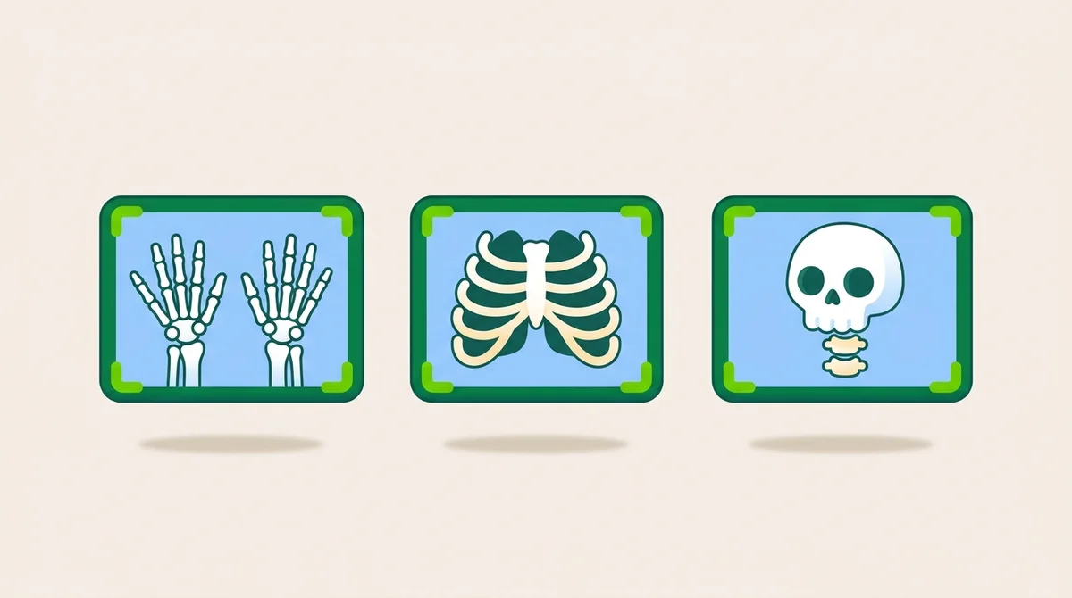
ARRT Procedures is the largest single domain on the Radiography Boards by chapter count. Combined Extremity (~11%) + Thorax/Abdomen (~8%) + Head/Spine/Pelvis (~7%) = roughly 30% of the exam, or 60 questions out of 200.
Procedures rewards a different kind of memorization than physics or patient care. You don’t have to memorize every projection ever named (there are hundreds in the radiography literature). You have to know the workhorses cold, the standard projections you’ll use 90% of the time in the clinic, and recognize them in question stems.
This is the master guide.
The positioning vocabulary
Before any specific projection, the ARRT assumes fluency in the language.
The anatomic position: standing erect, palms forward, feet pointing ahead. Universal reference for all directional terminology.
Body planes:
- Sagittal: divides left/right (midsagittal divides into equal halves)
- Coronal: divides anterior/posterior (also called frontal)
- Transverse: divides superior/inferior (also called axial)
Projections (path of the central ray through the body):
- AP, anterior to posterior
- PA, posterior to anterior
- Lateral, side
- Oblique, angled
Positions (how the body is placed):
- Supine, face up
- Prone, face down
- Recumbent, lying down (any position)
- Erect, standing or sitting
- Decubitus, lying on side with horizontal beam
Decubitus modifiers are tested heavily:
- Left lateral decubitus, patient lying on left side, beam horizontal
- Right lateral decubitus, patient lying on right side, beam horizontal
- Dorsal decubitus, supine, beam horizontal (lateral image)
- Ventral decubitus, prone, beam horizontal (lateral image)
The chest workhorses
Chest radiography is the most-ordered exam in medicine. The ARRT tests it accordingly.
PA chest (the standard): 72-inch SID, patient erect, chin extended, arms back to clear the scapulae, full inspiration. At least 10 posterior ribs above the diaphragm indicates adequate inspiration.
Lateral chest: left lateral by default (places the heart closer to the IR, less magnification). Arms raised over the head. True lateral, no rotation.
Lordotic AP: patient leans back so the central ray is angled relative to the body (or angle the CR cephalic 15–20°). Demonstrates apices and middle lobe free of clavicular superimposition.
Lateral decubitus: patient on the affected side for free fluid, opposite side for free air. Horizontal beam to demonstrate small pleural effusions or pneumothorax not visible on the upright PA.
The extremity workhorses
Hand: PA, PA oblique (45° lateral rotation), lateral.
Wrist: PA, lateral, PA oblique (45° lateral rotation). For suspected scaphoid fracture, add the Stecher (PA with ulnar deviation, central ray 20° proximally toward the elbow). The Stecher elongates the scaphoid and reveals fractures missed on routine PA.
Elbow: AP, internal oblique, external oblique, lateral. When the patient cannot fully extend, take two AP projections, one for the humerus, one for the forearm, per trauma protocol.
Shoulder: AP internal rotation + AP external rotation for routine. Grashey (AP oblique 35–45° body rotation toward affected side) for the glenohumeral joint in profile. Trauma: scapular Y or transthoracic lateral.
Foot: AP axial (10° posteriorly toward the heel), AP oblique (medial 30–40° rotation), lateral.
Ankle: AP, AP mortise (15–20° internal rotation to open the talocrural joint), lateral.
Knee: AP, lateral (5–7° cephalic angle), AP oblique. For the intercondylar fossa: Holmblad (PA with 70° flexion) or Camp-Coventry (PA with 40–50° flexion + 40–50° caudad CR). Patellofemoral joint: Settegast (sunrise).
Hip: AP pelvis with 15–20° internal rotation of legs, AP hip with 15–20° internal rotation, Danelius-Miller cross-table axiolateral for trauma.
The spine workhorses
Cervical spine routine: AP open-mouth (odontoid view), AP axial (CR 15–20° cephalic), lateral, obliques (45° rotation, CR 15–20° cephalic for posterior obliques).
Open-mouth odontoid demonstrates C1, C2, and the dens between the upper teeth and skull base. Patient’s mouth open, beam perpendicular.
Cervical trauma: cross-table lateral first, every time, before any other view. Don’t lift the head until the C-spine is cleared.
Thoracic spine: AP (perpendicular CR), lateral (with breathing technique to blur the ribs).
Lumbar spine: AP, lateral, oblique (45° rotation). The posterior oblique demonstrates the side closest to the IR. The Scotty Dog pattern is the pars interarticularis, a “collar” indicates spondylolysis.
L5-S1 spot: 30° caudad CR for women, 35° caudad for men, centered 1.5” inferior to the iliac crest.
Sternum: RAO 15–20° (throws sternum off the spine), lateral.
Ribs: above the diaphragm on inspiration; below on expiration.
The skull workhorses
The ARRT loves skull projections. Memorize the workhorses by name + central-ray angulation + cranial baseline.
Three baselines you must know:
- OML (orbitomeatal line), outer canthus to EAM
- IOML (infraorbitomeatal line), infraorbital margin to EAM, 7° below OML
- GAL (glabellomeatal line), 7° above OML
Caldwell PA: CR 15° caudad to OML. Frontal bone, anterior ethmoid sinuses, superior orbital fissures.
Towne AP axial: CR 30° caudad to OML (or 37° to IOML). Occipital bone, dorsum sellae projected within foramen magnum.
Waters parietoacanthial: CR perpendicular, OML 37° to the IR. Maxillary sinuses, orbital floors free of petrous ridge superimposition.
SMV (submentovertical): IOML parallel to IR, CR perpendicular to IOML. Skull base, sphenoid sinuses, mandibular condyles.
Schueller axiolateral TMJ: CR 25–30° caudad. Closed and open mouth comparison.
Stenvers PA petrous: 45° head rotation. Demonstrates petrous portion in profile.
Paranasal sinus rule (a single rule that wins many questions)
The patient must be erect and the central ray must be horizontal. Both are required to demonstrate air-fluid levels. Recumbent imaging or angled beams obscure the diagnostic finding.
If a question describes a sinus exam with a recumbent patient, the right answer is always to discuss with the radiologist before substituting recumbent views.
Trauma adaptive positioning
The “Don’t Move Rule”: in trauma, move the tube and the IR, never the patient or the injured part. Adapt the projection, not the anatomy.
For suspected hip fracture: do not abduct or internally rotate. Use the Danelius-Miller cross-table axiolateral.
For suspected C-spine injury: cross-table lateral first, every time.
For unstable forearm: take two AP projections (one for humerus, one for radius/ulna) rather than forcing rotation.
When an ordered projection violates the Don’t Move Rule, document why a substitute view was performed and which radiologist authorized it.
Pelvis: the 15–20° rule
For AP pelvis, internally rotate the legs 15–20°. This compensates for femoral anteversion and opens up the femoral neck for visualization. The same rotation applies to the AP hip.
Do not internally rotate when fracture is suspected, use the cross-table axiolateral instead.
Common fractures by name
| Fracture | Location |
|---|---|
| Colles | Distal radius, dorsal displacement (fall on outstretched hand) |
| Smith | Distal radius, volar displacement |
| Boxer’s | 5th metacarpal neck (punching) |
| Bennett | 1st metacarpal base |
| Scaphoid | Most commonly fractured carpal bone (avascular necrosis risk) |
| Monteggia | Proximal ulna + radial head dislocation |
| Galeazzi | Distal radius + distal radioulnar joint dislocation |
| Pott | Bimalleolar ankle |
| Jones | 5th metatarsal base (transverse fracture) |
| Lisfranc | Tarsometatarsal complex |
| Salter-Harris | Pediatric epiphyseal (Types I–V) |
How the ARRT tests Procedures
Across roughly 60 questions in this domain:
- 18–22 extremity (hand, wrist, elbow, shoulder, foot, ankle, knee, hip)
- 14–18 thorax/abdomen (chest, abdomen, GI, urography)
- 12–16 head/spine/pelvis (skull, sinus, spine, pelvis)
- 6–8 trauma adaptive positioning
- 4–6 fracture identification
The pattern: scenario describes the clinical question, the patient situation, the ordered exam. Your job is to identify the projection that demonstrates the diagnostic finding without making the injury worse.
Study path for Procedures
- Positioning Foundations, vocabulary, planes, projections
- Upper Extremity, hand to shoulder
- Lower Extremity, foot to hip
- Trauma and Adaptive Positioning
- Axial Skeleton: Spine and Bony Thorax
- Skull, Face and TMJ
- Paranasal Sinuses
- Chest Radiography
- GI and Hepatobiliary Procedures
- Special Procedures: CNS and Circulatory
Then drill all three procedures categories: Extremity (105 questions), Thorax/Abdomen (75), and Head/Spine/Pelvis (68).
The mental model that works
When you read a procedures question, run this mental sequence:
- What anatomy is being imaged? (chest, hand, knee, skull, spine, etc.)
- What is the clinical question? (fracture? infection? air-fluid level? joint space?)
- Is this trauma? (if yes, Don’t Move Rule applies)
- What’s the workhorse projection for this combination?
- What’s the central-ray angulation and patient rotation?
- What special view, if any, demonstrates the specific clinical question? (Stecher for scaphoid, Holmblad for intercondylar fossa, Grashey for glenohumeral joint)
Run that sequence reflexively and you have all the procedures questions in 90 seconds each.
Start studying
Sign up free, the first chapter (Foundations of Practice) and 50 practice questions. The Premium subscription unlocks all 11 chapters of procedures content and the 248 procedures practice questions across the 3 categories.